|
|
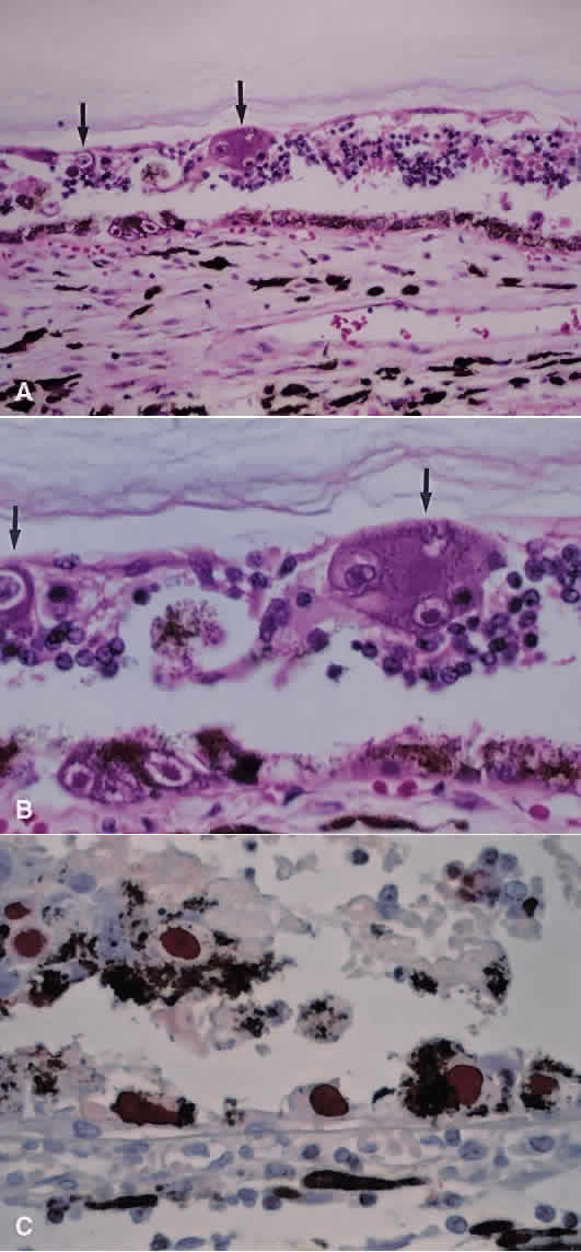
| Fig. 11. A and B. Photomicrographs of retina from a patient with cytomegalovirus retinitis showing retinal atrophy and characteristic large (megalo-) cells (arrows) with intracytoplasmic and intranuclear inclusion bodies (hematoxylin and eosin; A × 200, B × 500). C. Immunoperoxidase stain for cytomegalovirus antigen shows presence of virus (indicated by rust color) within cells in the retina, including the retinal pigment epithelium (immunoperoxidase; × 500). |