|
|
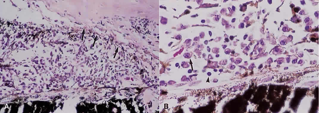
| Fig. 2. A. Photomicrograph of iris root and anterior chamber angle. There is apposition of iris to trabecular meshwork with closure of the angle by peripheral anterior synechiae (arrows) (hematoxylin and eosin; × 200). B. Chronic inflammatory cells including plasma cells (arrow) and lymphocytes (arrowhead) can be seen within the iris stroma (hematoxylin and eosin; × 500). |