|
|
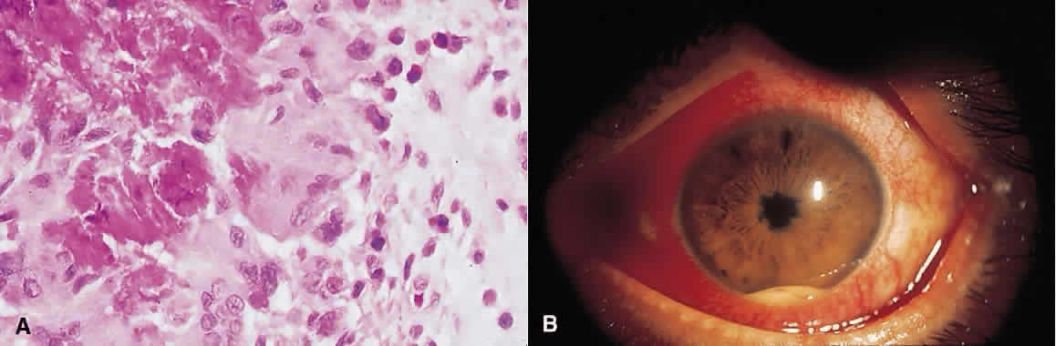
| Fig. 1. A. Photomicrograph of conjunctival biopsy specimen from patient with parasitic infection. Eosinophils are visible in the upper right corner of the image. Red-pink material at left of image represents the Splendore-Hoeppli phenomenon seen in such infections (hematoxylin and eosin; × 500). B. HLA-B27-associated acute anterior uveitis. Intense conjunctival injection, posterior synechiae, and hypopyon with admixed hemorrhage can be seen. |