|
|
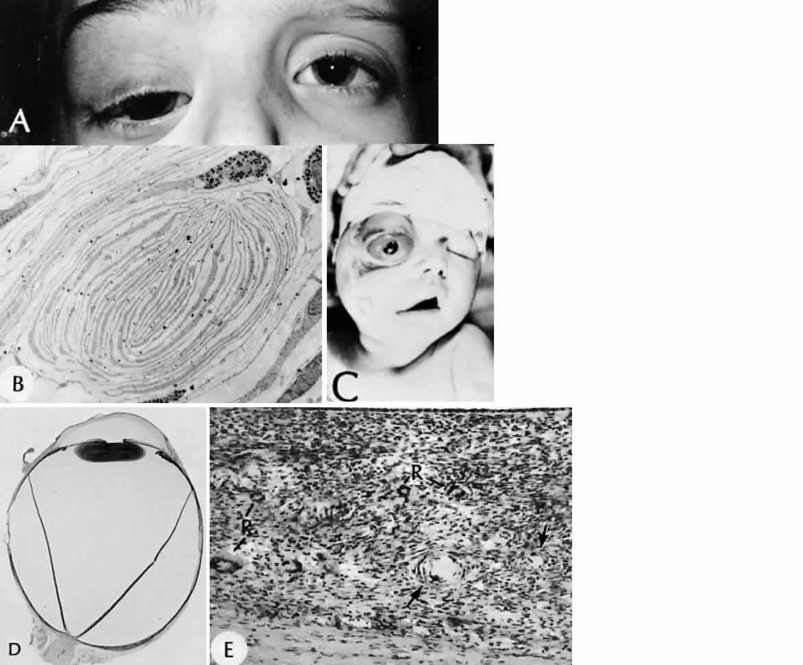
| Fig. 8. Neurofibromatosis. A. Plexiform neurofibroma of the right upper eyelid obscures most of an enlarged, blind glaucomatous eye. B. Ovoid body in a hamartomatously thickened choroid reveals the concentric lamellae of Schwann cell processes (× 3,000). C. Child born with neurofibromatosis (SEI 73–113). D. Whole eye removed at autopsy from the child shown in A (H&E, × 3 [SEI 73–191]). Note the diffuse thickening of the choroid posteriorly. E. High magnification of a diffuse choroidal hamartoma composed of structures resembling tactile nerve endings (arrows), rosette formation (R), and cells resembling nevus cells (H&E, × 101 [SEI 73–247]. (A and B, courtesy of Dr. RC Eagle Jr; C, D, and E, courtesy of Dr. L Calkins) |