|
|
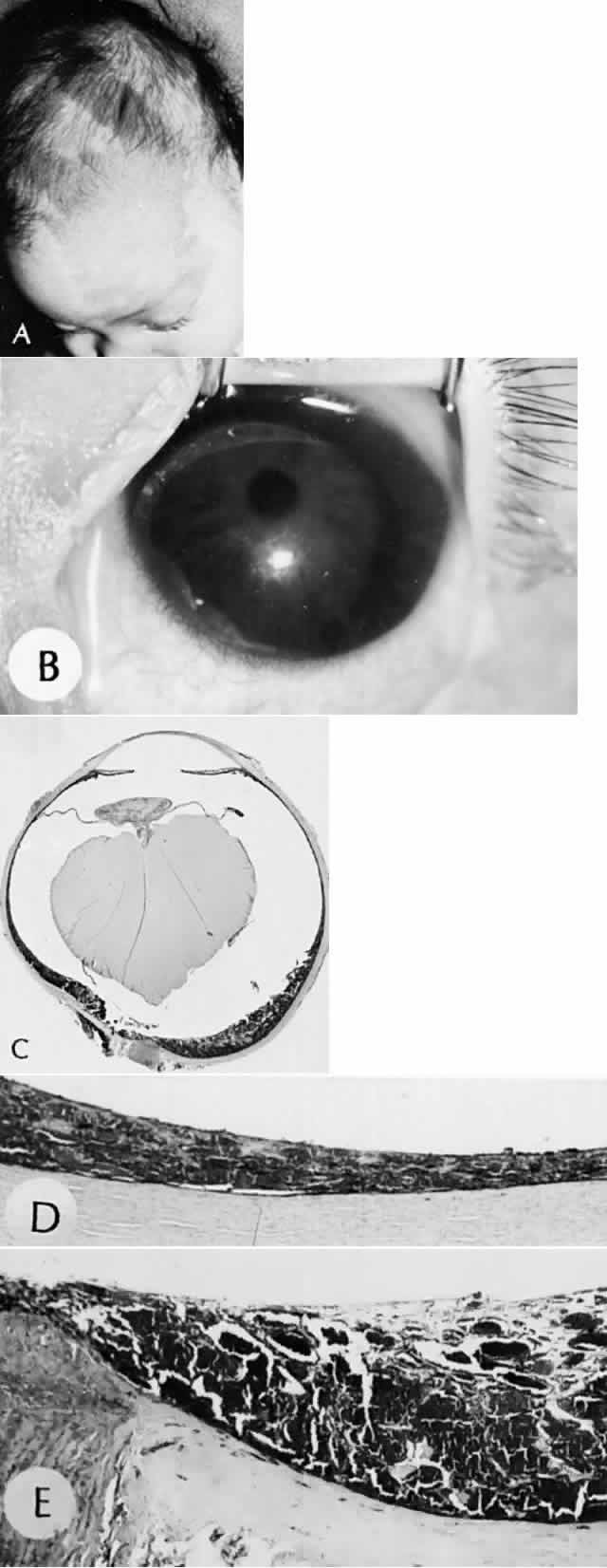
| Fig. 5. Meningocutaneous angiomatosis. A. Facial nevus flammeus (port-wine stain) along the distribution of the first division of the trigeminal nerve in an infant. B. Left eye in the same patient shows an enlarged, cloudy cornea caused by congenital glaucoma. C. Choroid is thickened posteriorly by a cavernous hemangioma that blends imperceptibly into the normal choroid, as shown in D. E. Cavernous hemangioma of the choroid in the same eye shows large, thin-walled, blood-filled spaces. (A and B, clinical [SEI 79-22 and 79-23]; C, H&E, × 5 [SEI 73–187]; D and E, H&E, × 16 [SEI 79-25 and 79-74]. A and B, courtesy of Dr. HG Scheie; C, D, and E, courtesy of Dr. R. Cordero-Moreno) |