|
|
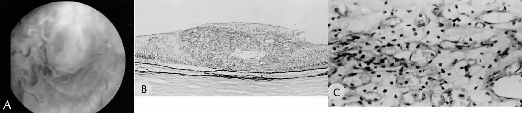
| Fig. 3. Angiomatosis of the retina. A. Fundus picture of retinal angioma in a 16-year-old patient. B. Hemangioblastoma (capillary hemangioma) replaces the full thickness of the retina. C. High magnification shows capillary blood-filled spaces intimately associated with characteristic pale, foamy, polygonal stromal (astrocytic) cells.(A, fundus picture [SEI 79-21]; B, H&E, × 40 [SEI 73–207]; C, H&E, × 252 [SEI 73–209]. Courtesy of Dr. DH Nicholson). |