|
|
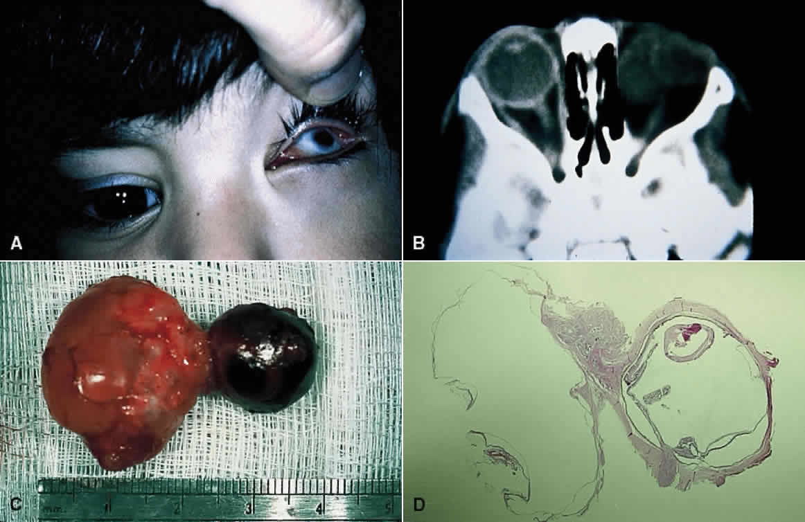
| Fig. 1. Microphthalmia with cyst. A. The microphthalmic left eye in a 2-year-old girl. B. Computed tomographic scan showing the cyst nasal to the microphthalmic eye. C. Gross specimen, showing that the cyst is larger than the eye. D. Low-power photomicrograph showing the relatively well-formed eye and large cyst (HE, × 1). There is focal retinal dysplasia. (UTHSC-SA EP 169. Courtesy of Charles R. Leone, MD) |