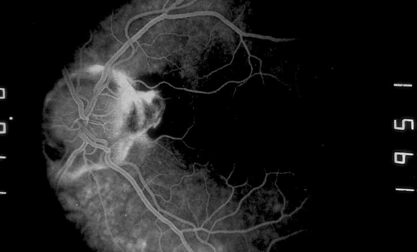
Fig. 38
Presumed ocular histoplasmosis. Fluorescein angiography shows a small peri-papillary choroidal neovascular membrane.