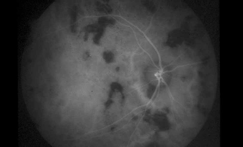
Fig. 29
Serpiginous choroiditis. ICG angiography later frame shows multiple hypofluorescent spots in the location of the lesions seen on color photography.