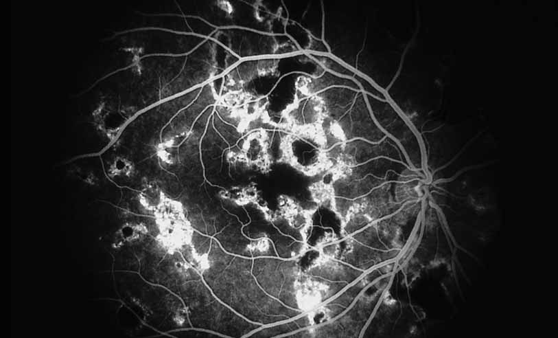
Fig. 27
Serpiginous choroiditis. Fluorescein angiography shows areas of hypofluorescence and hyperfluorescent staining at lesion edges.