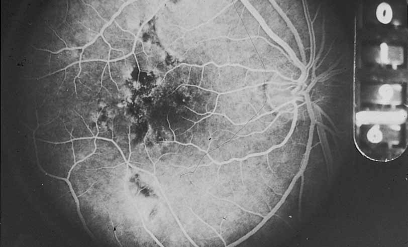
Fig. 25
Acute multifocal placoid pigment epitheliopathy. An early frame of the angiogram in
Figure 26
discloses areas of hypofluorescence corresponding to the ophthalmoscopically visible lesions (Courtesy of Joseph Michaelson).