|
|
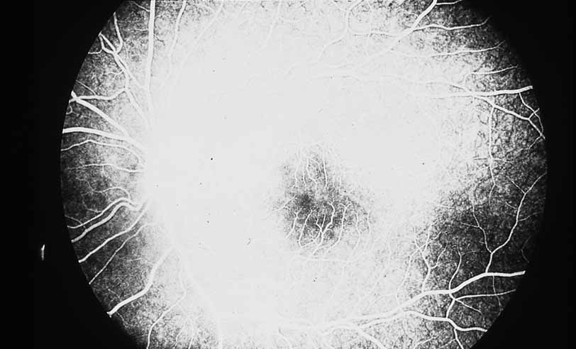
| Fig. 5 Luetic neuroretinitis. A late frame of the angiogram in Figure 4 shows extensive staining of the entire area corresponding to leakage into the outer retina and staining of the pigment epithelium. The patient was successfully treated with intravenous penicillin therapy and subsequently developed diffuse “leopard spot” changes in the pigment epithelium characteristic of resolved luetic neuroretinitis. |