|
|
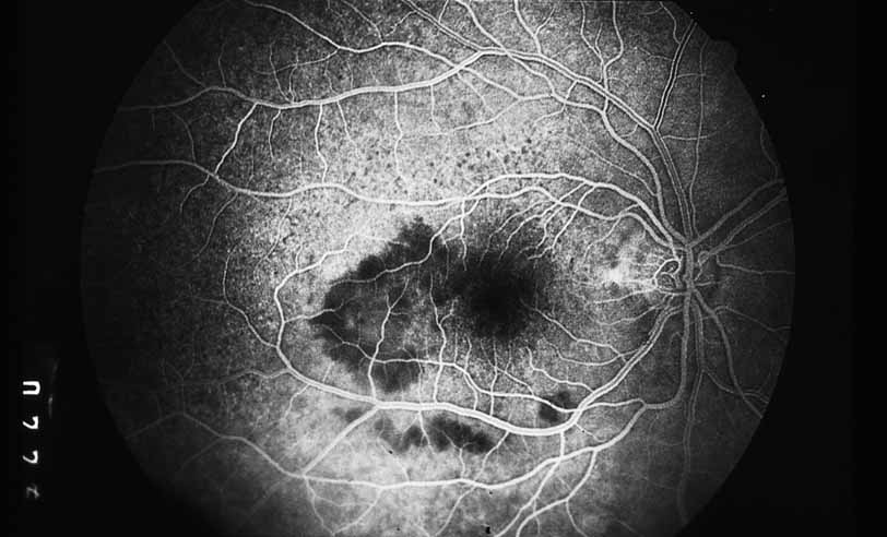
| Fig. 3 Luetic neuroretinitis. Early-frame angiogram of eye of middle-aged man who presented with vision loss and a peculiar yellow---green discoloration of the outer retina and pigment epithelium. The patient was not known to be immunosuppressed, and subsequent serologic testing confirmed the presence of a positive VDRL and FTA-ABS. The early frame of the angiogram demonstrates a broad area of hypofluorescence corresponding to the clinical lesion. Note the subtle punctate leopard spots above the lesion. |