|
|
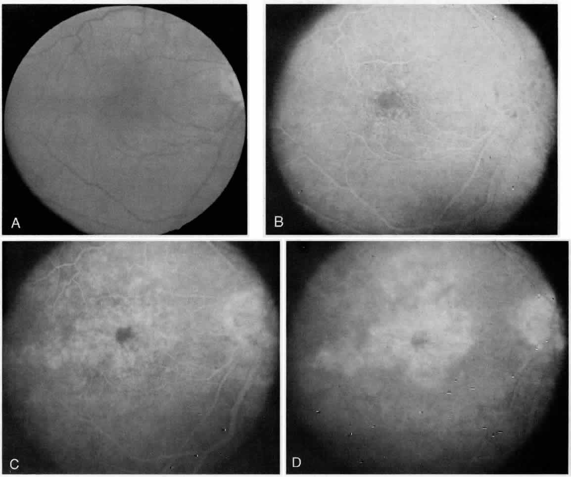
| Fig. 6. Case 6 is a 65-year-old woman with a 5-year history of non-insulin dependent diabetes and was seen visual acuity of 20/80 in her right eye 5 months following uncomplicated extracapsular cataract extraction and intraocular implant. Clinical examination revealed cystic thickening of the macula without evidence of diabetic retinopathy (A; Color Fig. 6). A fluorescein angiogram was obtained, which confirmed the diagnosis of psuedophakic cystoid edema. The early frames of the angiogram revealed an intact FAZ (B); as the study progressed, uniform leakage was noted (C) with the petalloid appearence of dye accumulation (D) seen in the latest frame. Note also the late staining of the optic disc. Comment: The differntial between psuedophakic or aphakic cystoid macular edema and diabetic macular edema is often difficult despite fluorescein angiography. The angiogram shown here is characteristic of classic psuedophakic cystoid macular edema. |