|
|
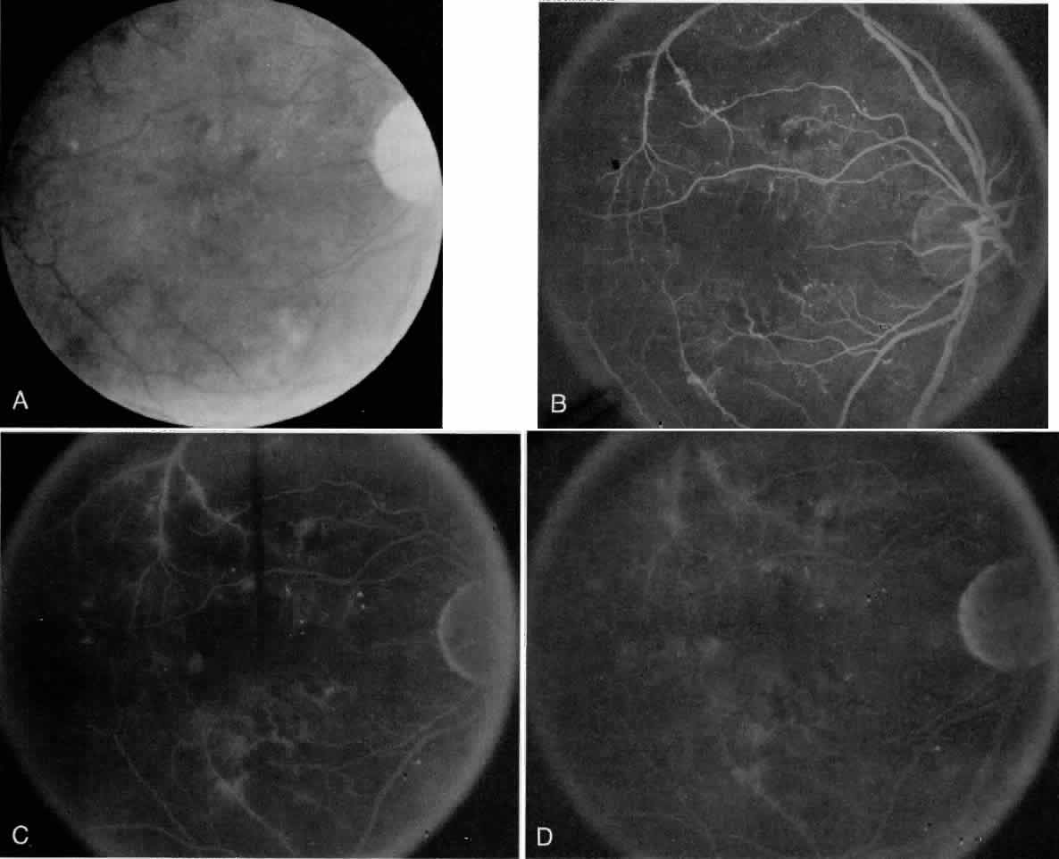
| Fig. 1. Case 1 is a 60-year old man with a 20-year history of noninsulin dependent diabetes and chronic open-angle glaucoma controlled by topical medications who complained of decreased vision in his right eye. Visual acuity was 20/100. Minimal background retinopathy, intraretinal hemorrhages inferior to the fovea, and macular thickening, with an enlarged cup-to-disk ratio are noted on dialated fundus examination with visual loss out of proportion to his clinical examination (A; Color Fig. 1). Fluorescein angiography was therefore obtained, revealing marked irregularity of the capillary-free zone and perifoveal nonperfusion (B). There is no evidence of central leakage in the mid- and late frames (C, D). His visual loss can be explained by his macular ischemia secondary to diabetic retinopathy. Comment: This is an example of a patient with unexplained visual loss, out of proportion to the clinical examination; a fluorescein angiogram is indicated in this situation. |