|
|
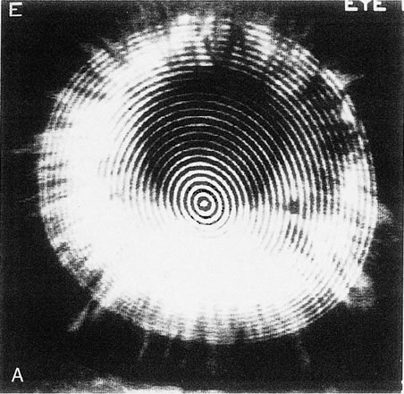
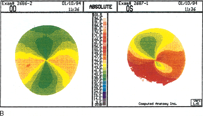
| Fig. 12. Keratoglobus. A. Videokeratoscopic image of a patient with keratoglobus in the left eye showing inferonasal narrowing of the rings, indicating steepening, but without the pear-shaped configuration seen in pellucid marginal degeneration. B. Videokeratography of the right eye shows marked against-the-rule astigmatism of 6.3 diopters (D). C. The left eye shows irregular astigmatism with quite irregular power distribution. The axis of lowest corneal power is shifted about 35 degrees from the vertical axis, with a very asymmetric bow-tie configuration and with the inferior low-power semimeridian positioned above an area of high power at the inferior peripheral cornea. This area of peripheral inferior corneal steepening extends to the steep oblique semimeridians. (Karabatsas CH, Cook SD: Topographic analysis in pellucid marginal degeneration and keratoglobus. Eye 10:451–455, 1996.) |