|
|
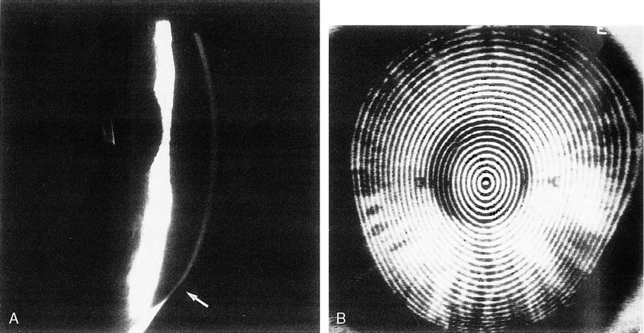
| Fig. 11. Pellucid marginal degeneration (PMD). A. Slit topography showing inferior corneal thinning (arrow) 1 to 2 mm from the limbus, extending from the 5- to 8-o'clock positions in both eyes. B. Videokeratoscopic image shows a typical pear-shaped image with compression of the inferior rings. C. Corneal topographic maps (absolute scale) showing against-the-rule astigmatism of 10.6 diopters (D) in the right eye. The left eye shows enantiomorphic symmetry (mirror image) to the right eye with 11.9 D of against-the-rule astigmatism. In early PMD the power of the cornea is least at a vertical axis very close to 90 degrees. The area of greater power is presented in a bow-tie configuration of two semimeridians inferior and oblique to the horizontal axis. (Karabatsas CH, Cook SD: Topographic analysis in pellucid marginal degeneration and keratoglobus. Eye 10:451–455, 1996.) |