|
|
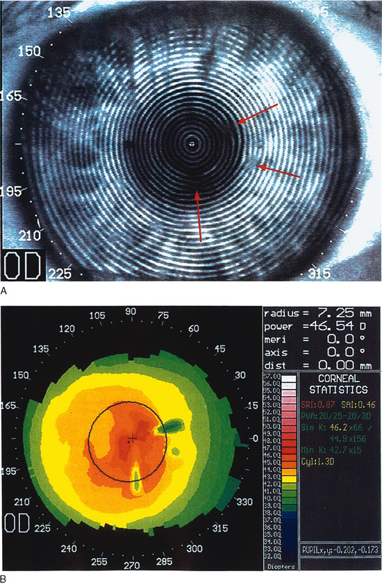
| Fig. 8. Recurrent corneal erosion. In this patient with recurrent corneal erosion, no epithelial abnormality could be detected on biomicroscopic examination between attacks. A. The videokeratograph showed irregularity of the mires (arrows) in the 10-degree, 270-degree, and 350-degree semimeridians, about 1 to 2 mm from the corneal center. B. These correspond to areas of focal flattening on the topography. (Corbett MC, Rosen ES, O'Brart DPS: Corneal Topography Principles and Applications. London, BMJ Books, 1999.) |