|
|
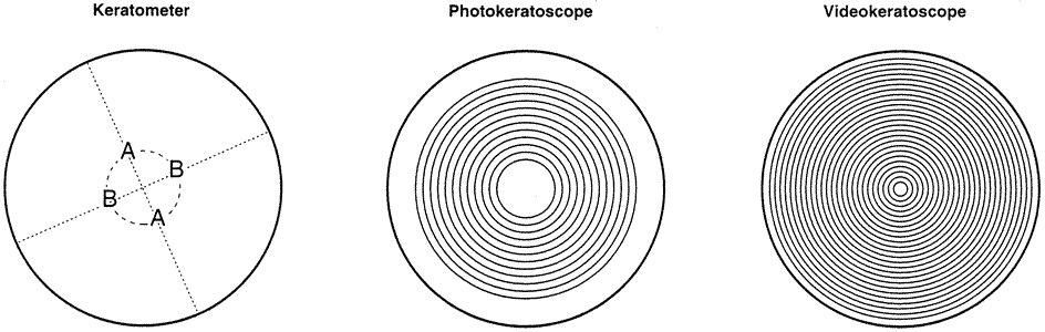
| Fig. 2. Mires. Representation of the corneal area covered by the mires of the keratometer (two perpendicular pairs of mires, A and B, situated on an anulus approximately 3 mm in diameter), photokeratoscope (12 rings), and computer-assisted videokeratoscope (25 rings). (Corbett MC et al: Corneal Topography Principles and Applications. London, BMJ Books, 1999.) |