|
|
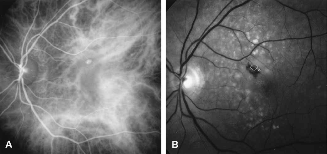
| Fig. 7 A, Mid-phase indocyanine green (ICG) angiogram demonstrates a focal spot of hyperfluorescence representing an area of intraretinal neovascularization in a patient with retinal angiomatous proliferation. Identification of this lesion would be difficult with ICG study alone. The software capabilities of the digitized system can be used to allow this image to be warped and overlayed over a red-free image. B, A tracing of the outline of the hyperfluorescent lesion can be superimposed on the red-free photograph. This overlay tracing provides good landmarks to permit accurate localization if photocoagulation is performed. |