|
|
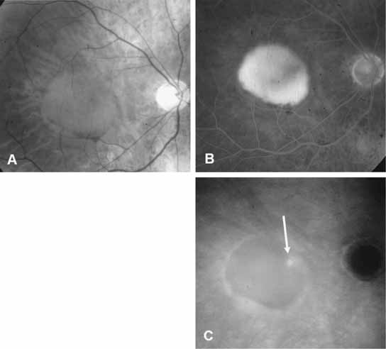
| Fig. 4 A, The red-free photograph of a patient with neovascular age-related macular degeneration shows a large pigment epithelium detachment (PED) in the central macula. B, Late-phase fluorescein angiogram demonstrates hyperfluorescence of the serous PED. No focal area of choroidal neovascularization can be identified. C, Late-phase indocyanine green (ICG) angiogram reveals a focal spot of hyperfluorescence (arrow) representing an area of localized choroidal neovascularization. The ICG molecule, which is 98% protein-bound, does not leak from the neovascular membrane, and the PED remains relatively hypofluorescent throughout the study. |