|
|
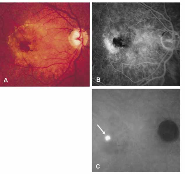
| Fig. 2 A, Clinical photograph demonstrates subretinal and intraretinal hemorrhages as well as detachment of the retinal pigment epithelium and the neurosensory retina in a patient with neovascular age-related macular degeneration. B, Late-phase fluorescein angiogram reveals blocked fluorescence from the hemorrhages and indistinct leakage. C, A late-phase ICG angiogram demonstrates a well-defined hyperfluorescence or so-called focal hot spot (arrow) representing a retinal angiomatous proliferation. This lesion is well visualized through the area of hemorrhage because of good penetration of the infrared light used in ICG angiography. |