|
|
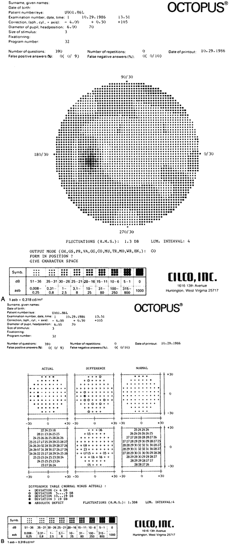
| Fig. 5. A. A cursory review of the gray-scale printout of this Octopus Perimeter 30° full threshold examination of the central field demonstrates no gross abnormalities in this 34-year-old ocular hypertensive. B. Inspection of the numeric printout of the differences (center, bottom) between actually determined threshold sensitivities (left, bottom) and those expected (right, bottom) reveals several points of minimally reduced sensitivities in the inferior Bjerrum area. These findings are suggestive of the development of early visual field defects and warrant close scrutiny on future testing. Note the use of symbols in the difference display (top, center) using the difference table as a key (bottom). |