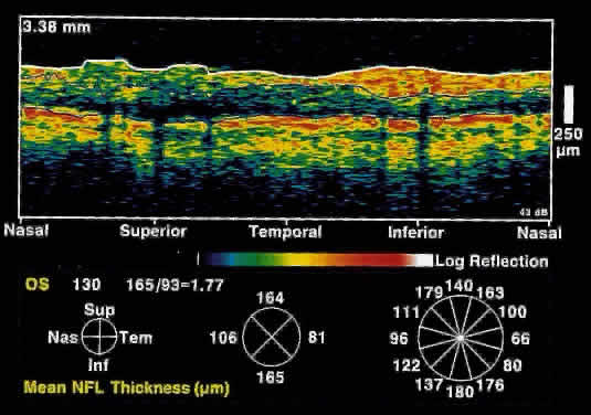
Fig. 16.
Circular OCT image of a normal eye taken in cylindrical section around the optic nerve head. Note the thicker nerve fiber layer superiorly and inferiorly.