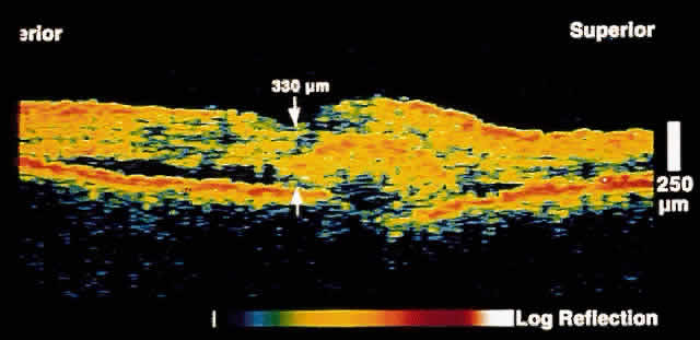
Fig. 10.
OCT image through the fovea of a patient with a choroidal neovascular membrane secondary to age-related macular degeneration. The neovascular tissue appears to have penetrated Bruch's membrane to lie primarily in the subretinal space.