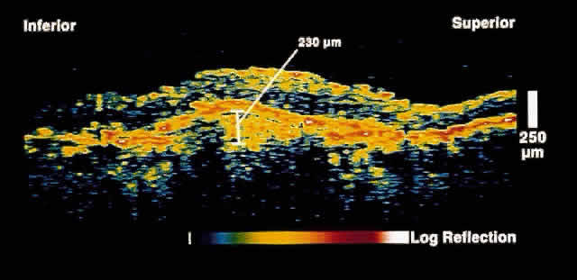
Fig. 9.
OCT image through the fovea of a patient with a fibrovascular RPE detachment secondary to age-related macular degeneration. Note the moderately reflective layer throughout the sub-RPE space, which corresponds to fibrovascular tissue.