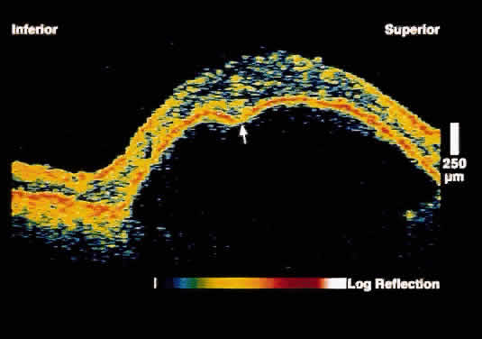
Fig. 7.
OCT image through the fovea of a patient with a serous RPE detachment secondary to age-related macular degeneration. Note the sharp contrast between the posterior border of the detached RPE band and the underlying serous fluid.