|
|
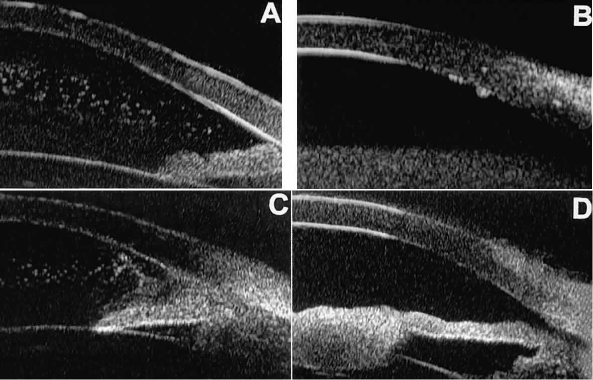
| Fig. 20. UBM features of uveitis. A. Inflammatory cells suspended in anterior chamber aqueous. B. Keratic precipitates appear as small sonoreflective bumps on peripheral corneal endothelium. C. Hypopyon. Mass of inflammatory cells fills inferior anterior chamber angle, and dispersed cells are suspended in central anterior chamber aqueous. D. Inflammatory mass of pupillary zone of iris. Mass disappeared after corticosteroid therapy. |