|
|
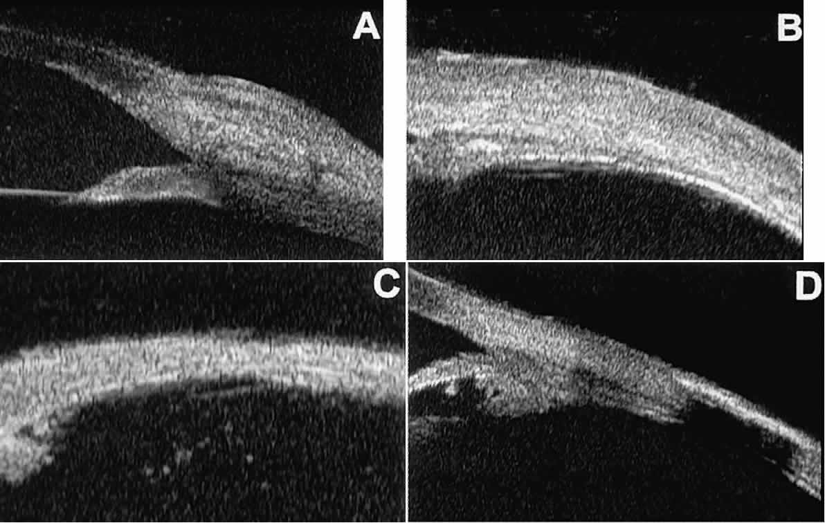
| Fig. 16. UBM features of anterior scleral disorders. A. Nodular anterior scleritis appears as fusiform thickening of limbal sclera. Note apparent lamellae of heterogeneous reflectivity within region of thickening. B. Diffuse anterior scleritis appears as nonfocal scleral thickening in region of inflammation. C. Scleral thinning subsequent to necrotizing anterior scleritis. Note underlying vitreous cells. D. Scleral hyaline plaque appears as dense, hyper-reflective plate several millimeters from horizontal limbus; dense lesion “shadows” deeper tissues. |