|
|
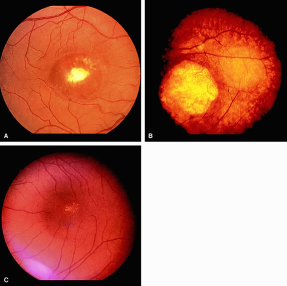
| Fig. 5. Three different patients with Best's disease showing the remarkable range of changes that can occur. A. Subretinal neovascular membrane. B. Atrophic central retinal pigment epithelium (RPE) loss and several soft extramacular vitelliform lesions. C. Punctate drusen-like changes in foveal-parafoveal area. |