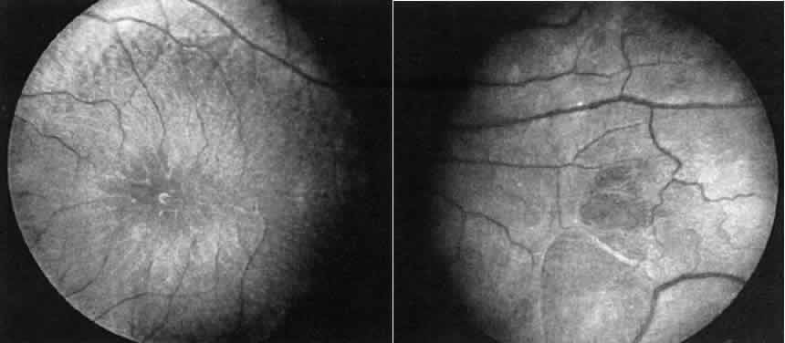
Fig. 17.
Case 14. See text for details.
Left.
Macular area.
Right.
Peripheral retina of another patient with X-linked retinoschisis showing the diaphanous “veils” or inner layer retinoschisis.