|
|
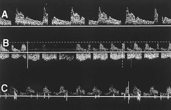
| Fig. 4. Velocity waveforms obtained using color Doppler imaging of a healthy eye. A. The ophthalmic artery (OA) shows steep systolic peaks that slope down to much lower end-diastolic velocities. B. The central retinal artery (CRA) and central retinal vein (CRV) waveforms are recorded simultaneously. The CRA waveform is similar to that of the OA but less steep. The CRV waveform shows significantly less pulsatility than the arterial waveforms. C. The posterior ciliary artery (PCA) waveform is similar to that of the CRA. |