|
|
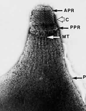
| Fig. 2. Electron micrograph of the apical complex of a T gondii tachyzoite. Subpellicular structures have been revealed by negative staining with phosphotungstic acid (× 82,000). APR, anterior polar ring; C, conoid; PPR, posterior polar ring; MT, microtubule; P, remnant of pellicle. |