|
|
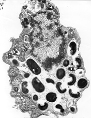
| Fig. 2. Electron micrograph of a eukaryotic host cell that contains Encephalitozoon intestinalis spores and developing forms inside a septated parasitophorous vacuole. Reprinted from the Parasite Image Library of the Centers for Disease Control and Prevention Website on Laboratory Identification of Parasites of Public Health Concern (DPDx). (Available at http://www.dpd.cdc.gov/dpdx/HTML/ImageLibrary/Microsporidiosis_il.htm accessed February 17, 2005.) |