|
|
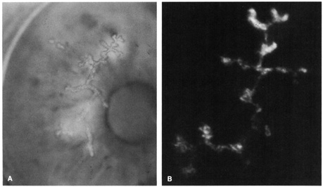
| Fig. 2. A: Herpes simplex dendritic epithelial keratitis stained with fluorescein. B: Impression cytology of the superior portion of this epithelial dendrite stained with an immunofluorescent technique to identify herpes simplex virus type 1. (Reprinted with permission from Simon MW, Miller D, Pflugfelder SC, et al: Comparison of immunocytology to tissue culture for diagnosis of presumed herpesvirus dendritic epithelial keratitis. Ophthalmology 99:1408, 1992.) |