|
|
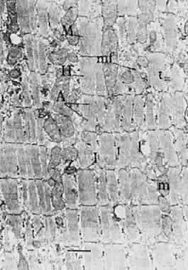
| Fig. 21. Low-power electron micrograph of longitudinal section of fibrillar type fiber. Myofibrils are well delineated by intervening sarcoplasm. Even at this low magnification, myofibrils are seen to be made up of myofilaments. Triads of T system are seen en face and in cross section. Also present are mitochondria, glycogen particles, and lipid droplets. A, A band; e, extracellular space; f, thin myofilament (actin); ff, thick myofilament (myosin); g, glycogen granule; H, H band; I, I band. Glutaraldehydeosmium fixation, original magnification × 14,000. (Kroll AJ: Neuroophthalmology 3:38, 1967.) |