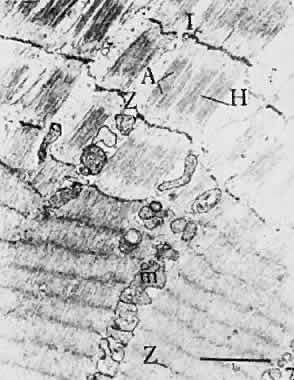
Fig. 19.
Electron micrograph of muscle fibers unstretched (
bottom
), and stretched (
top
). Note separation of Z-line and difference in width of I-band (I). A, A-band; H, H-zone. (Kroll AJ: Neuroophthalmology 3:42, 1967.)