|
|
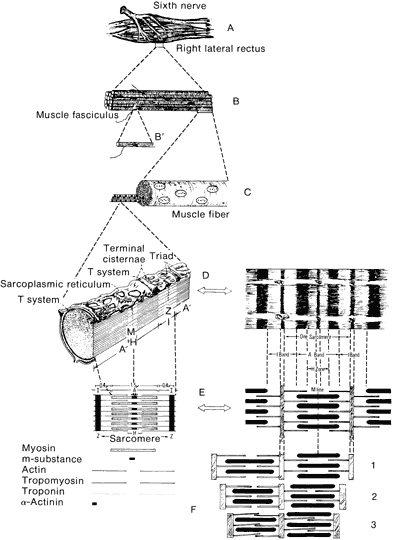
| Fig. 18. A schematic dissection of an extraocular muscle with gradually increasing magnification of the component parts. A. Right lateral rectus muscle viewed from the medial side showing distribution of the sixth cranial nerve over the medial surface of the muscle. B. A muscle fiber composed of many fasciculi, one of which is shown enlarged in B' with a small motor nerve on its surface. A single muscle fiber (C) is composed of myofibrils that can be seen extending from the left cut surface of the fiber. One myofibril is enlarged (D) to indicate the various component parts and bands. The left portion of D is an artist's representation of the fibril with surrounding T system, the terminal cisternae, and the combination of these into triads. In the right portion of D is an artist's representation of a typical photomicrograph showing the various components. E, Artist's drawing of the various parts of the fibril and the relationship one to another as shown more clearly in E, right. F. Schematic of how the various portions exist and the relaxed form: (1) partially contracted, (2) strongly contracted, and (3) maximally contracted state. |