|
|
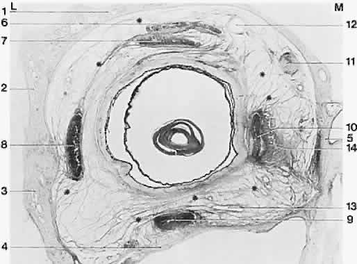
| Fig. 12. Frontally sectioned histologic section (60 degree) of an adult right orbit, at a level in the orbit 3.2 mm anteriorly from the posterior pole of the eye. Asterisk, connective tissue septa; 1, frontal bone; 2, greater wing of sphenoid; 3, zygomatic bone; 4, maxilla; 5, ethmoid; 6, superior levator palpebrae muscle; 7, superior rectus muscle; 8, lateral rectus muscle; 9, inferior rectus muscle; 10, medial rectus muscle; 11, superior oblique muscle; 12, superior ophthalmic vein; 13, branches of inferior ophthalmic vein; 14, medial check ligament. Notice size difference of muscles; intermuscular membrane is well developed here. M, medial; L, lateral. Acidic fuchsin-picrin acid/van Gieson stain, original magnification × 2.5. (Koornef L: Arch Ophthalmol 95:1271, 1977.) |