|
|
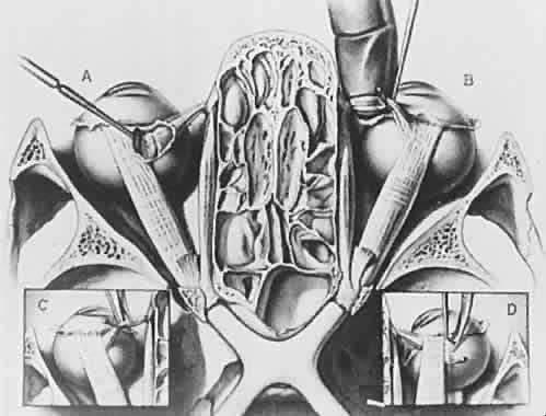
| Fig. 1. A coronal section through the human skull just below the level of the orbital roof. The conjunctiva, eyelids, orbital fat, and blood vessels have been removed. Contents of the infratemporal fossa have also been removed. Note that the orbits are separated by ethmoid air cells and that the medial walls of the orbit are parallel. The lateral walls diverge one from another at an angle of approximately 90 degrees and an orbital axis drawn as a center line through the orbit would have an angle of about 23 degrees. The left portion of the figure, A, shows the sheath of superior oblique muscle opened and the tendon partially divided. The retractor is partially drawing out the superior rectus muscle. The levator palpebrae has been removed. In the right portion of the illustration labeled B, the tendon of superior oblique muscle has been caught with a muscle hook and a gloved finger has been placed beneath the tendon. Note the trochlea arising from the superior portion of the medial orbital wall. (C) below, left, shows the reflected tendon of superior oblique muscle beneath a muscle hook and about to be divided by surgical scissors. Another approach to dividing the tendon is shown in D, right inset, with the insertion of the muscle outlined by the muscle hook and scissors coming from the anterior direction about to cut the muscle free from the globe. The optic nerves can be seen passing through the optic canal in the posterior central portion of the illustration forming the chiasm. The pituitary gland has been removed from the pituitary fossa in this illustration. (Berke RN: Trans Am Ophthalmol Soc 44:314, 1946). |