|
|
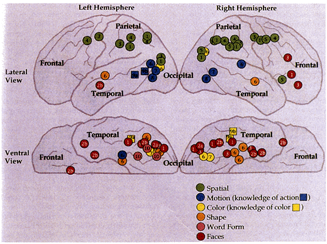
| Fig. 16. Human visual processing streams identified in PET imaging studies. Top. Lateral views of the left and right hemispheres. Bottom. Ventral views of the hemispheres. Numbers in the symbols indicate the study reporting each activated focus of increased blood flow. In some instances, multiple nearby foci of activation are shown as a single focus, representing their center of gravity. Study numbers are as follows: 1, face and location matching-to-sample; 2a, gender discrimination; 2b, face identify; 3, working memory for faces and locations; 4, shifting attention to spatial locations; 5, spatial working memory; 6, selective attention to color, shape, and velocity; 7, passive perception of color and motion; 8, passive perception of motion; 9a, word generation of object attributes from line drawings of objects; 9b, word generation of object attributes from object words; and 10, perception of word forms. (Adapted from Ungerleider LG: Functional brain imaging studies of cortical mechanisms for memory. Science 270:769, 1995) |