|
|
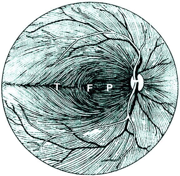
| Fig. 1. Course of the retinal ganglion cell axons within the nerve fiber layer of the retina. (F, fovea; P, papillomacular bundle; T, temporal raphe). (Redrawn from Kline LB. Optic Nerve Disorders. Ophthalmology Monographs No. 10. San Francisco: American Academy of Ophthalmology, 1996:4) |