|
|
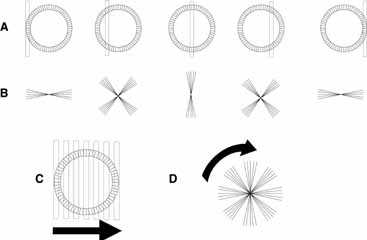
| Fig. 2 When a narrow (1 mm or less) stenopeic slit is positioned in front of the pupil, a lenticular halo is visualized (lines A and B). The visible portion of the halo formed (line B) depends on the orientation of the lens fibers in the regions of the pupil that are illuminated (line A). When the stenopeic slit is moved sequentially across the pupil (C), a lenticular halo that resembles the rotating arms on a windmill will become evident (D). |