|
|
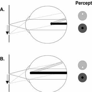
| Fig. 1 The use of relative entoptic parallax to estimate the position of an opacity in the eye. In the two cases illustrated here, a pinhole source is shifted downward from the optical axis (as indicated by the arrow), and the amount and direction of the displacement of the shadow of an opacity relative to the entoptic image of the pupil is evaluated. In both cases (A and B), the shadow of the opacity (black shadow) appears centered within the entoptic image of the pupil (red lines) when the pinhole is positioned on the optic axis. In the first case (A), the opacity is positioned posterior to the entrance pupil of the eye. When the pinhole is shifted downward, both the entoptic image of the pupil (green lines) and the shadow of the opacity (yellow shadow) are displaced upward, but there is relatively less displacement of the shadow. As a result, the shadow of the opacity is no longer centered in the entoptic image of the pupil. This is perceived as if the shadow moved in the opposite direction from the image of the pupil. The closer the opacity lies to the retina, the greater the relative displacement. In the second case (B), the opacity is positioned anterior to the entrance pupil of the eye. Once again when the pinhole is shifted downward, both the entoptic image of the pupil (green lines) and the shadow of the opacity (yellow shadow) are displaced upward, but in this case there is relatively more displacement of the shadow. As a result, the shadow of the opacity is no longer centered in the entoptic image of the pupil. This is perceived as if the shadow moved in the same direction as the image of the pupil. The closer the opacity lies to the pinhole, the greater the relative displacement. |