|
|
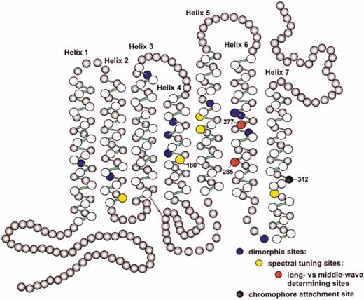
| Fig. 26 A schematic representation of the L\/M opsin molecule. Each circle is an amino acid. The colored amino acids represent those sites that are known to differ among X-linked cone opsins. Positions 227 and 285 are sites that together shift the spectral peak of a pigment approximately 20 nm. They are the determinants of whether a pigment is classified as L or M. Other sites produce smaller shifts in the wavelength of peak sensitivity. Some dimorphic sites do not appear to shift peak sensitivity in all pigments. Site 180 plays an important role in variations in normal color vision. (Reprinted form Neitz M, Neitz J. Molecular genetics and the biological basis of color vision. In Backhaus WGK, Kliegl R, Werner JS (eds): Color Vision: perspectives from different disciplines. Berlin: Walter de Gruyter, 1998:101–119. Copyright 1998. Reproduced with permission of Walter de Gruyter Gmbtl Co. KG.) |