|
|
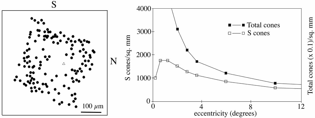
| Fig. 5 A: Locations of individual S cones in human retina. The center of the foveola is indicated by a triangle. The central 100μm is devoid of S cones. S, superior; N, nasal. B: Distributions of S cones and of L plus M cones across the retina. The peak density of S cones is at approximately 1 to 2 degrees eccentricity. The cone density scale for L and M cones is compressed by a factor of 10 relative to the scale for S cones. (Redrawn from Curcio CA, Allen KA, Sloan KR, Lerea CL, Hurley JB, Klock IB, Milam AH: Distribution and morphology of human cone photoreceptors stained with anti-blue opsin. J Comparative Neurol 312: 610-624, 1991. Copyright © 1991 Wiley-Liss, Inc., A Wiley Company. Reproduced with permission of John Wiley and Sons, Inc.) |