|
|
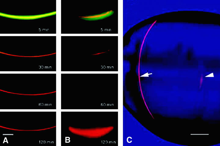
| Fig. 13. Results from a study in which a relatively small fluorescent solute, carboxyfluorescein (CF) and a large, 40 kDa solute, tetramethylrhodamine dextran (TD) were injected into a lens fiber cell. After 120 minutes, CF was observed to diffuse away from the injected fiber into neighboring cells (A), whereas the larger solute, TD, was retained (B). When a similar injected is made into a fiber cell deep in the lens core, both CF and TD diffuse from the injected cell; this is seen in panel C in which TD is clearly visible 3 hours after injection into a peripheral fiber (arrow) but has mostly diffused away from the injection site in a core fiber (arrowhead). The results demonstrate the ability of solutes to pass from fiber to fiber via gap junctions and, in the lens core, via regions of cell-cell fusion. (Shestopalov VI, Bassnett S: Expression of autofluorescent proteins reveals a novel protein permeable pathway between cells in the lens core. J Cell Sci 113:1913, 2000) |