|
|
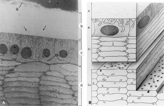
| Fig. 9. A. Light micrograph of the zonular fibers (a), capsule (b), lens epithelium (c), and cortex (d) approximately 0.7 mm anterior to the equator of the lens. The zonules insert into the most superficial portion of the capsule. The capsule contains scattered linear densities (arrows) because of the presence of coarse fibrils. The epithelial cells are tall and columnar; their base faces the capsule, and their apex faces the lens cortex. The round nuclei are either central in the cytoplasm or displaced slightly toward the apex. Elaborate basal infoldings of the cells membranes are apparent. The Golgi zone appears as a dense accumulation in the apical cytoplasm (e).The rectangular or hexagonal cells of the lens cortex are seen in cross section. They have a fairly homogeneous cytoplasm. Interlocking processes occur along both the short and the long sides of the hexagon in the superficial layers of the cortical cells. In deeper cortical cells the interlocking processes are found chiefly along the long sides of the hexagons (× 1300). B. 3-D drawing of the lens indicating the interrelation of the capsule and underlying lens cells. The capsule (a) shows inclusions of fine filamentous material. The anterior lens epithelium (b) shows interdigitation of its basal surface with adjacent cells. The superficial cells of the cortex show the hexagonal shape and interdigitations at their hexagonal ends, as well as along their edges (c1, arrows). The deeper cortical cells (c2) also show a tongue-and-groove type of interdigitation along their long sides (arrows), but the interlocking is absent at the short ends. (Hogan MJ, Alvarado JA, Weddell JE: Histology of the Human Eye. Philadelphia, WB Saunders, 1971) |