|
|
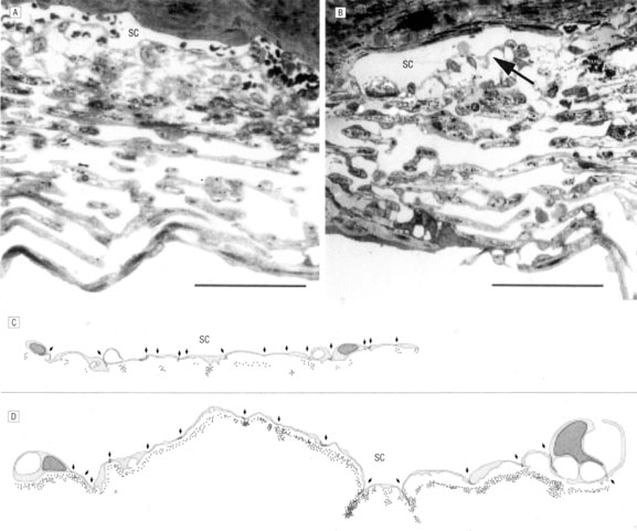
| Fig. 21. H-7 inhibits myosin light chain kinase and ρ kinase to block cellular actomyosin-driven contractility; leading to rapid deterioration of actin-containing stress fibers and focal contacts. A and B: Light micrographs (bars indicate 50 μm) of trabecular meshwork and Schlemm's canal in monkey eyes treated with H-7 ((1-[5-isoquinoline sulfonyl]-2-methyl piperazine), 300 μmol/L (B) or vehicle (A). The juxtacanalicular area (arrow in B) and intercellular spaces are extended, and extracellular material is lost. C and D: Schematic drawings depicting 15-cell stretches (cell–cell junctions marked by arrows) along the Schlemm's canal (SC) of control (C) and H-7–treated (D) eyes showing distribution of perfused gold particles the in juxtacanalicular area. The location of individual gold particles is represented by dots. Expanded areas are available for fluid drainage in H-7 treated eyes. (From Sabanay I, Gabelt BT, Tian B, et al: H-7 effects on structure and fluid conductance of monkey trabecular meshwork. Arch Ophthalmol 118:955, 2000, with permission.) |