|
|
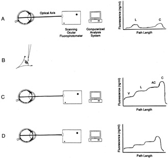
| Fig. 10. Principles of measurement of aqueous flow by ocular fluorophotometry. A: Optical axis of eye is scanned for background fluorescence with a scanning ocular fluorophotometer. B: Topical application of drops of fluorophore (2% fluorescein) applied to cornea. C: After a suitable delay (approximately 15 hours), to allow fluorescein to diffuse from the corneal depot to the aqueous humor, the eye is scanned once again. D: Repeated scans at 30-minute to 1-hour intervals over a 3- to 6-hour period facilitate monitoring of decline in fluorescence of aqueous humor with time. This can be related mathematically to aqueous flow rate (a calculation often performed by computer) after subtraction of background fluorescence and derivation of anterior chamber volume from keratometry and pachymetry determinations. The graphs to the right of the diagrams indicate typical fluorescence patterns obtained along the optical axis at each stage in the procedure. C, cornea; AC, anterior chamber; L, lens; V, vitreous. |